Parkinson's Patient care guide for Families
Complete Parkinson's home care guide for families, rehabilitation, safety and daily living.
Read MoreThis resource page aims to help family members, caregivers to support a loved one after stroke discharge. It covers daily care, feeding and swallowing, mobility, medicines, vital signs, rehabilitation, communication, skin care, continence, nutrition, infection prevention, and managing conditions like hypertension or diabetes. Includes step-by-step instructions, checklists, recovery timelines, and emergency guidance. Advice follows trusted global stroke guidelines and should be individualized to each patient’s needs.
Choose bathing time appropriately. Best time to bath is when the patient has the most energy; gather all supplies beforehand; never leave them unattended in the bath.
For bedbound patients, perform a bed bath in sections: cover the body with towels, wash and dry one area at a time, re-cover to keep the patient warm and maintain privacy. Use mild, fragrance-free soaps and rinse thoroughly. Apply a gentle, alcohol-free lotion (e.g. petroleum-based moisturizer) after bathing to prevent dry skin.
Check skin (especially bony areas like heels, elbows, hips, tailbone) daily for redness or sores; document and consult with your physician if any concerns.
Turn or reposition the patient every 2 hours during the day (at least every 4 hours at night) to prevent pressure ulcers (also known as bed sores). Use pillows/ foam wedges between knees and ankles, and under elbows and heels, to evenly distribute pressure. Keep sheets smooth and flat (wrinkles can rub and cause sores). When turning, use a draw sheet or lift device if available, and employ safe body mechanics: bend knees, keep back straight, and slide rather than lift to avoid shear. The caregiver should always explain each move to the patient, encouraging participation as much as possible.
The following video explains essential care tips for bedsores (pressure sores), focusing on safe cleaning and wound management. Learn why using normal saline is better than antiseptics for preventing infection and how to manage large wounds with loose dressing. Perfect for caregivers, nurses, and healthcare professionals, this guide highlights practical wound care techniques to improve healing, comfort, and patient recovery in everyday caregiving situations.
Brush teeth (or dentures) twice daily with fluoride toothpaste. Include tongue brushing to remove bacteria. Offer frequent sips of water or mouthwash rinse (if allowed) to keep the mouth moist. If the patient has a Ryle’s/ Nasogastric (NG) tube or low saliva, apply lip balm for cracked lips. Wipe saliva before meals or if secretions build up. (Note: If oral suction is needed, please take help of trained personal and equipment.)
Inspect the skin daily, especially under tape or medical devices (tubes, dressings). Use lotion to keep dry areas healthy. Change bed linens and patient clothing when damp with sweat or urine to reduce infection risk. Avoid harsh soaps, detergents, or lotions with perfumes/ alcohol.
Dress the patient in loose, comfortable clothing with front-fastening and elastic waists. Use slipper or velcro shoes to eliminate risks of tripping. Undress the affected (weak) limbs last and dress them first to avoid strain on stronger side.
Clean the nostril site daily with warm water; apply water-soluble lubricant to prevent nose irritation. Secure tape/ straps on face to avoid accidental dislodgement. Flush the NG tube with 30 – 60 ml warm water before and after any feeding or medication. For the tube, mark the external length and check regularly to ensure it hasn’t migrated. Report pain or resistance when flushing – it may indicate blockage or position change.
Patients recovering from stroke may try to pull out a Ryle’s tube (nasogastric feeding tube) because of throat/nasal irritation, confusion, restlessness, discomfort, delirium, or impaired judgment after stroke. Families should know this is common and preventable, but it also needs prompt attention.
To reduce the chances of a patient pulling out a Ryle’s tube, first make sure the tube is comfortable and properly secured. Check regularly for nasal irritation, throat dryness, loose tape, tugging, or blockage, as discomfort often causes patients to touch or remove it. Keep extra tubing neatly looped and fixed to clothing so it does not hang or pull. Calmly remind the patient why the tube is needed, especially if they are confused after stroke. Maintain a quiet, familiar environment, ensure adequate sleep, and keep glasses or hearing aids on if used, as confusion increases agitation. During high-risk times such as nighttime or repositioning, supervise more closely. Keep the patient’s hands occupied with a towel or cushion for distraction. If repeated attempts continue, inform the treating doctor or nurse so the cause can be assessed and safer solutions considered.
Dysphagia (difficulty swallowing) affects over half of stroke survivors. To reduce aspiration risk: have the patient sit upright (90°) during and after meals (remain upright ≥30 min). Use chin-tuck posture if advised by therapist. Remove distractions (TV, phone) to promote focus on eating. Offer small bites and sips, and ensure thorough chewing and swallowing before the next bite. Encourage the patient to cough and clear their throat after swallowing to expel any material in the airway. If the patient drools or coughs often while eating, notify the therapist; maintain mouth hygiene and suction as needed.
Follow speech-therapist or dietitian recommendations. For moderate swallowing issues, modify textures: puree or mash solid foods (dal, soups, mashed potatoes, curd, boneless fish, avoid chicken initially as the fibers can cause discomforts) and thicken liquids (honey-thick) to slow flow. Use the International Dysphagia Diet Standardisation Initiative (IDDSI) framework levels (0=thin, 1-slightly thick, 2-mildly thick, 3-liquidised solids, etc) as guidance. Avoid mixed textures (e.g. soups with chunks) and straws (which can speed liquids into the throat).
If prescribed, feed via the NG tube as follows (bolus or gravity method, per clinician instruction):
Provide a balanced diet as recommended. If eating orally, ensure high-protein, nutrient-dense foods (eggs, yogurt, lean meats) to aid healing.
Encourage the patient (with assistance) to move and change position in bed. Use rolling techniques: one person gently rolls the patient by shifting shoulders and hips, supporting the head. Place pillows under knees or heels and forearms for comfort. Teach log-roll technique (keeping spine aligned) when turning in bed to avoid twisting.
For patients who can sit up, instruct them to first roll onto one side, swing legs over the bed edge, and push up with arms to sit (the caregiver may assist by supporting the back or using a gait belt). Ensure feet are flat on the floor or use a footstool.
Always plan the transfer: remove obstacles, use gait belt if available, ensure footwear is non-slip. Sit the patient at bed edge, feet planted; ask them to lean forward and push up using stronger legs. The caregiver stands in front, knees bent, holds gait belt or torso. On cue, both rise, then the patient takes a few steps back until the chair touches their legs, then slowly sits. For wheelchair transfers, position the chair at 45° angle to the bed on the stronger side, and follow a similar pivot with support.
Use assistive devices as prescribed (walker, cane, wheelchair). Ensure devices are correctly sized and in good repair. Remove throw rugs and clutter; secure loose cords. Install night lights for bathroom trips. Always supervise initial ambulation and consider a transfer belt for added stability. Use chairs with arms for support during standing. Encourage appropriate footwear (non-slip socks or Velcro shoes).
Adapt the environment: install grab bars in bathroom, handrails in hallways, ramps or stair-lifts if needed. Keep frequently used items within easy reach to limit the need for bending or climbing. Use a firm mattress and chair to ease transfers. A raised toilet seat and bedside commode facilitate toileting.
Encourage daily range-of-motion (ROM) exercises to prevent joint contractures: gently bend/straighten elbows, wrists, fingers, hips, knees 5–10 times, 2–3 times a day. Ankle pumps (point/flex feet) prevent foot drop and clots. When able, incorporate gentle strength exercises (e.g. squeezing a soft ball, standing leg raises) under therapist guidance. Follow any prescribed home exercise plan (e.g. sit-to-stand practice, balance training, walking) to maximize recovery during the critical first 3–6 months.
Careful medication management prevents complications and stroke recurrence. Please follow the medication prescribed by your Doctor. If you face any specific question or discomfort please don't hesitate to reach out to your doctor for clarifications.
Keep a medication chart or use a medicine organizer with days/time slots. Administer meds at consistent times daily (e.g. morning aspirin with breakfast, evening statin at dinner). Set alarms or use smartphone apps for reminders. Maintain a copy of all prescriptions and dosages. Do not stop any medication abruptly unless instructed.
Measure daily (morning and evening) and keep a log. Targets: usually <140/90 mmHg (often <130/80). Inform doctor of readings consistently above or below targets.
If diabetic, check as instructed (e.g. fasting and postprandial), log values. Diet and medication adjustments aim to keep HbA1c near goal (~7% depending on guidelines).
Check pulse daily; irregular beats warrant medical attention. Use a simple palpation or home monitor. For patients with AF, monitor signs of clot (sudden limb pain/ swelling, chest pain).
Fever >100.4°F (38°C) may signal infection (pneumonia, UTI) and requires prompt evaluation. Ensure infectious causes are treated quickly.
Observe breathing; note any shortness of breath, cough, or signs of aspiration (wet voice, respiratory distress). Encourage deep breaths or use incentive spirometry post-stroke to prevent atelectasis.
For heart failure or renal issues, weigh daily and report sudden gain (fluid retention). For nutritional status, track weight weekly to catch malnutrition or fluid overload.
Note any new headache, weakness, numbness, vision changes, or speech difficulty; these could signify a recurrent stroke or complication. If any sudden focal neurological sign appears (e.g. one-sided droop, slurred speech, confusion), follow the FAST protocol and seek emergency care immediately.
Pleaes plan in advance for respones during any emergency. Keep all the phone numbers handy e.g. any neighbours, friends or relatives who can be present to support. Keep the ambulance, hospital emergency and your neurologists number handy. Also keep a list of current medication handy.
Initiate emergency steps in case you observe
If less severe but concerning (worsening weakness, fever >100.4°F, uncontrolled pain, increased confusion), consult with your doctor.
Continue exercises prescribed by therapists (e.g. ankle pumps, knee lifts, shoulder rolls). Aim for gradual increase in activity each day: frequent short walks (with assistance) versus few long walks. Use assistive devices as advised. Bed-bound patients should sit up to 30–60 minutes several times a day to improve circulation and breathing.
Focus on self-care tasks – allow the patient to do as much dressing, feeding, and grooming as able, using adaptive equipment (built-up utensils, sock aid, long-handled sponge). You may take help of an Occupational Therapist for inputs on home enviroment modifications. Encourage use of the weaker hand (with support from the unaffected) to maintain function. Practice fine motor skills: pick up coins, write, button/ unbutton. If hand spasticity develops, gentle stretching and speaking to therapist about splints is important.
Work on communication and swallowing exercises. For aphasia, use repetition games, naming objects, or picture cards. Encourage the patient to speak or gesture; be patient and attentive. For communication, use yes/ no questions, simple sentences, and allow extra response time. Consider communication boards or phones/ apps if recommended.
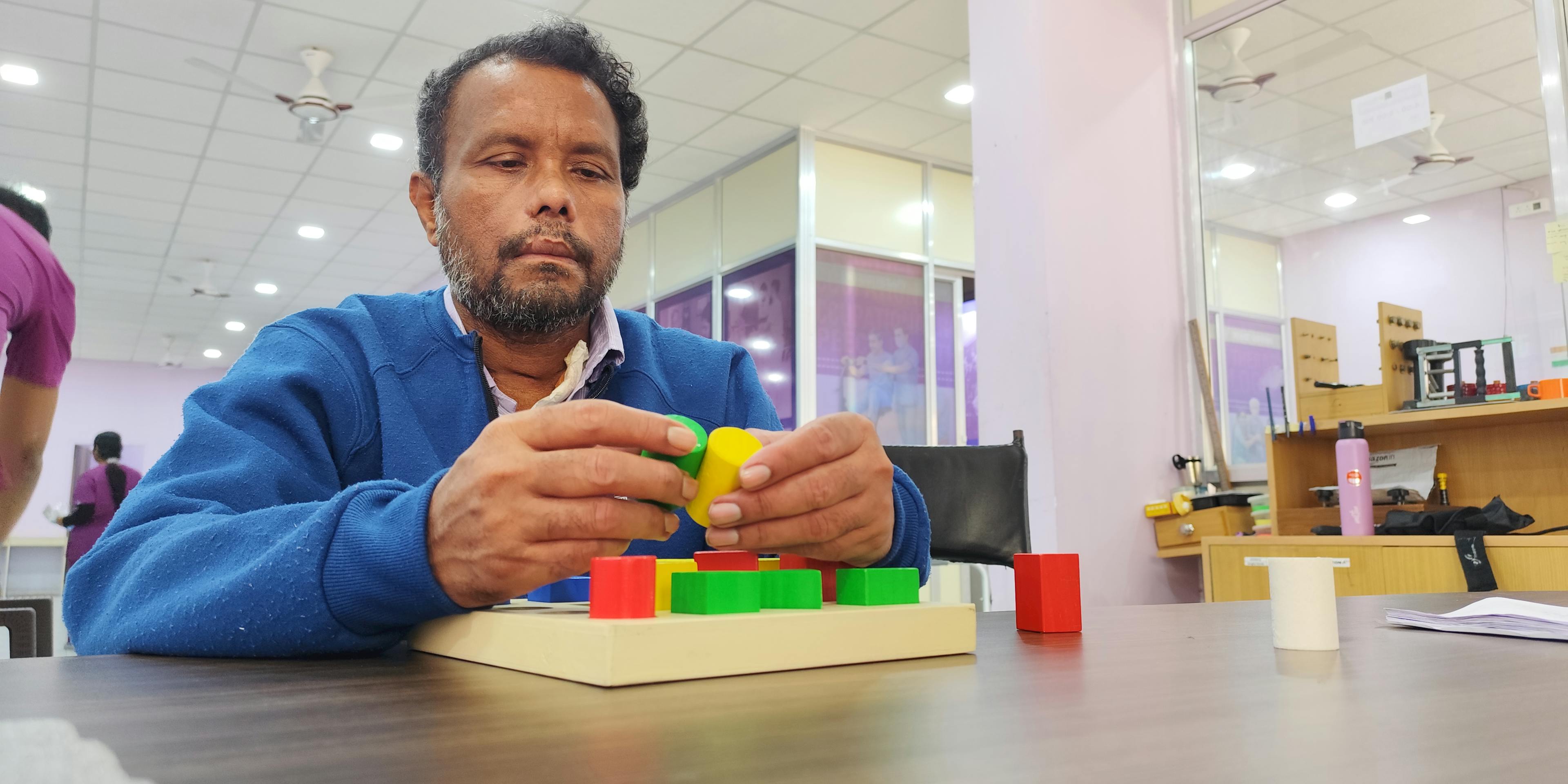
Engage in memory and problem-solving activities: simple puzzles, calendars (help re-orient date/ time), familiar music, or daily routine tasks. Establish a routine (consistent wake-up, meals, therapy times) to reduce confusion. Praise successes to boost confidence.
Recovery is most rapid in the first 3–4 months, so maximize therapy during this period. Goals should be reviewed with therapists regularly and adjusted as gains are made.
Speak clearly, slowly, using one idea per sentence. Use gestures, writing or drawing if possible. Confirm understanding by asking simple questions. Allow the patient to take time; avoid speaking over them. Implement communication cues (visual aids, gesture prompts). A speech therapist can teach specific strategies like “melodic intonation” or word-finding cues.
Post-stroke cognitive impairment may include attention deficits, memory loss, or executive dysfunction. Create a structured environment: use clocks, calendars, and routine schedules. Simplify tasks into steps and provide cues/ reminders (labels on cupboards, alarms for appointments). Ensure safety in decision-making; avoid multi-tasking. Encourage social interaction (visits, phone calls) to stimulate language and memory.
Be aware of post-stroke depression or frustration. Signs include tearfulness, apathy, or anger. Offer emotional support, listen empathetically, and consider professional psychological counseling if needed. Provide reassurance and involve family or a stroke support group for both patient and caregiver.
Pain is common after stroke and may begin immediately or develop gradually over the following weeks and months. Many family members think pain only comes from injury, but after stroke it can arise from several causes, including weak muscles, stiff joints, poor positioning, nerve changes, or medical complications. Pain can interfere with sleep, mood, confidence, movement, and participation in rehabilitation. Because some stroke survivors have speech, memory, or communication difficulties, they may not always explain their discomfort clearly. Families should watch for signs such as facial grimacing, irritability, guarding an arm or leg, refusing movement, restlessness, poor appetite, or disturbed sleep.
One of the most common causes of pain after stroke is shoulder pain. When the arm becomes weak or paralysed, the shoulder muscles may not support the joint properly. If the arm hangs unsupported, is pulled during transfers, or is moved roughly, pain and inflammation can develop. Reduced movement can also lead to stiffness in the shoulder joint over time.
Muscle and joint pain are also common because weakness often causes long periods of sitting or lying down. Joints that are not moved regularly may become stiff and uncomfortable. Muscles may tighten from poor posture or lack of stretching. This can cause pain in the neck, back, hips, knees, or ankles.
Some patients experience central post-stroke pain, which happens when the stroke affects areas of the brain that process sensation. In this case, the patient may feel burning, tingling, aching, electric shock-like sensations, or pain from light touch, even when there is no visible injury. This type of pain needs medical assessment.
Pain may also come from other medical conditions such as constipation, urinary infection, skin sores, swelling, arthritis, or previous injuries. Families should never assume all pain is simply part of recovery.
Spasticity is abnormal muscle tightness or stiffness caused by damage to the parts of the brain that control movement. After stroke, the normal signals that help muscles contract and relax smoothly may be disturbed. As a result, muscles can become overactive, tight, and resistant to movement. Spasticity commonly affects the weaker side of the body.
It may cause the shoulder to pull inward, the elbow to stay bent, the wrist to curl downward, the fingers to clench tightly into the palm, or the ankle to point downward. The leg may become stiff while walking, making movement difficult. Spasticity is different from true strength. A limb may feel strong because it is stiff, but the movement is poorly controlled.
The brain normally sends balanced messages to muscles, telling them when to tighten and when to relax. A stroke can damage these pathways, causing muscles to react too strongly to stretch or movement. Spasticity may appear days, weeks, or even months after the stroke.
It can become worse when the patient is in pain, tired, anxious, constipated, has an infection, or is moved suddenly. Poor positioning, skin irritation, and uncomfortable clothing or braces may also increase stiffness. Some patients notice more spasticity at certain times of day, especially in the morning or after poor sleep.
Family members may observe that the patient’s hand stays tightly closed, the arm remains bent against the body, or the leg becomes stiff during walking. Dressing and bathing may become harder because the joints resist movement. The patient may complain of pain during stretching or movement. Nails may dig into the palm if the hand remains clenched. Sudden jerking movements or muscle spasms can also occur. Reporting these signs early can help prevent worsening stiffness and loss of movement.
If pain and spasticity are not managed early, they can slow recovery and reduce quality of life. The patient may avoid therapy because movement hurts. Sleep can become poor, leading to fatigue and irritability. Walking, transfers, bathing, dressing, and toileting may become more difficult. Over time, joints may become fixed in bent positions, known as contractures, which are harder to treat. Tight skin folds and clenched hands may also lead to rashes, wounds, or infection. Early treatment often improves comfort, movement, and independence.
Physical Medicine and Rehabilitation specialists use several treatments to reduce spasticity and improve function. Regular stretching programs help keep muscles longer and joints more flexible. Range of motion exercises maintain mobility and reduce stiffness. These may be active exercises, where the patient helps move, or passive exercises, where a trained caregiver or therapist assists.
Positioning techniques and supportive splints may be used to keep joints aligned, prevent shortening, and improve comfort. Hand splints, ankle supports, or resting splints are commonly prescribed and should fit properly. In more severe cases, serial casting may be used to gradually stretch shortened muscles over time.
Electrical stimulation may be recommended in selected patients to activate weak muscles and improve movement patterns. Supported standing and weight-bearing programs can help posture, circulation, and muscle stretching. Gait training may be used for patients with leg stiffness to improve walking ability.
Doctors may also use botulinum toxin injections to relax specific overactive muscles for several months. This treatment is often combined with therapy for the best results. In some cases, oral medicines may be prescribed to reduce stiffness, although these must be monitored for side effects such as sleepiness or weakness.
Families should seek medical advice if the patient develops new severe pain, sudden swelling, redness, fever, rapid worsening of stiffness, repeated spasms that disturb sleep, skin wounds, shoulder injury after a fall or pulling, or a sudden decline in walking or transfers. These problems may need urgent treatment.
Sample Daily Care Checklist (must be personalized):
Pressure Ulcer Prevention: Turn schedule: e.g. change side at 8am, 10am, 12pm, etc. Use pillows. Document times.
Caring for a stroke survivor at home involves careful planning, teamwork with healthcare providers, and vigilant monitoring. This guide provides a non-medical caregiver with actionable instructions and safety-focused rationales across all domains of care. Always tailor the plan to the individual – their type of stroke, impairments, and home situation – and consult professionals for ongoing guidance. With structured care, patience, and support, caregivers can significantly aid recovery and quality of life for stroke survivors.
Caring for a family member after stroke can be physically tiring, emotionally draining, and mentally overwhelming. Many families try to manage everything on their own out of love and responsibility, but it is completely okay to ask for help. Seeking support is not a sign of failure—it is a sign of wisdom and care. Professional guidance from doctors, nurses, therapists, attendants, counsellors, or trained home-care teams can reduce stress, improve safety, and give you confidence in daily caregiving. When caregivers are supported, patients are supported better too. Taking breaks, sharing responsibilities, and protecting your own health are important parts of caring for your loved one.
A multidimensional neuro rehabilitation program can be a valuable step toward the best possible recovery. When different specialists work together with the family, the patient receives more complete and personalized care. Recovery may take time, but with the right team, steady effort, and family support, meaningful progress is always possible.
Knowledge That Guides Recovery
Get expert neuro rehabilitation tips from the doctors at Rehabana, Kolkata — simple guidance on stroke recovery, spinal cord injury rehab, Parkinson’s care, and enhancing quality of life after neurological conditions.
Complete Parkinson's home care guide for families, rehabilitation, safety and daily living.
Read More
SCI rehab guide for families after discharge. Skin care, mobility, bladder & bowel management, warning signs, recovery tips and safety steps.
Read More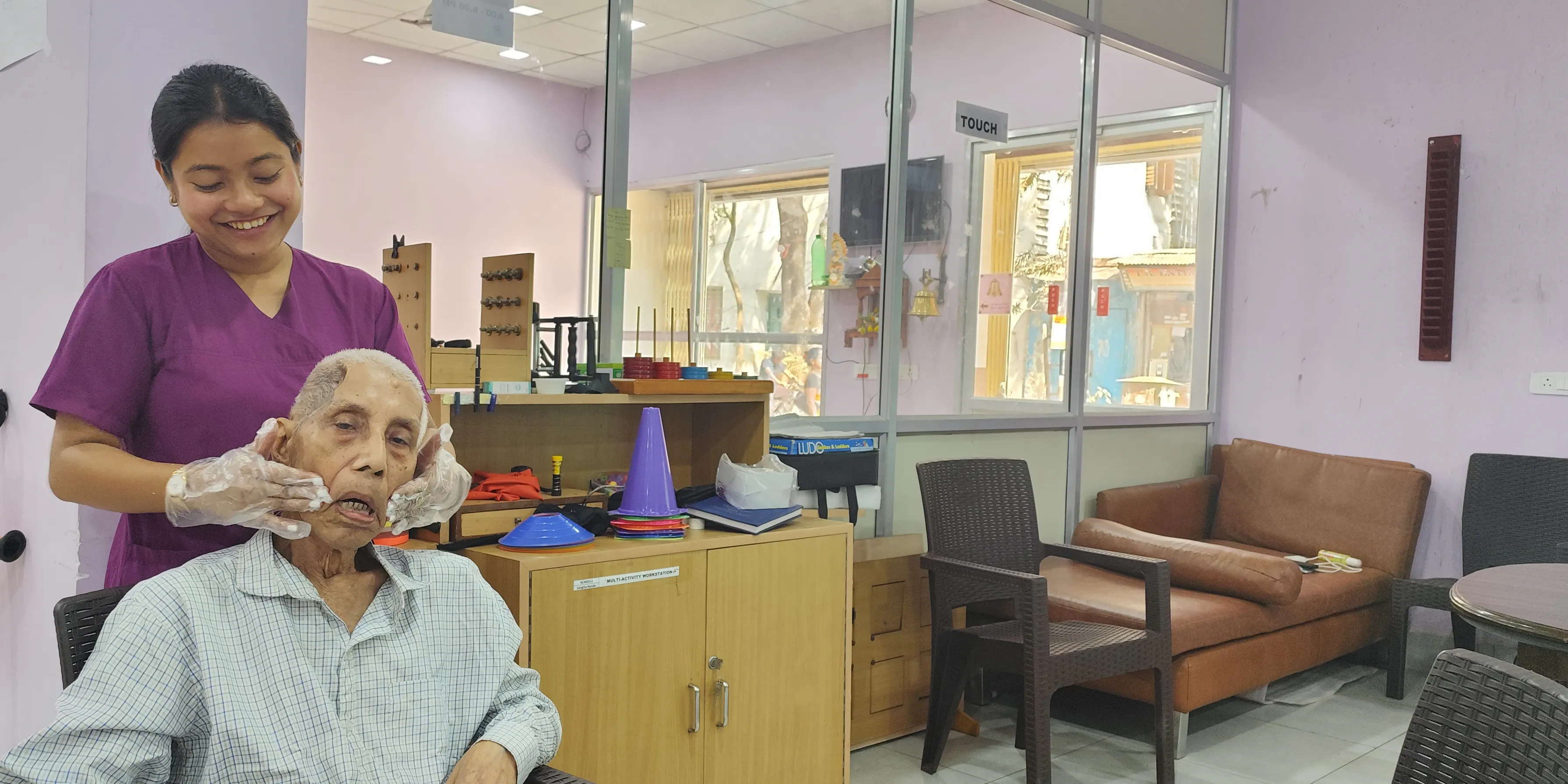
Stroke rehab guide for families after discharge. Daily support, mobility, feeding, medicines, warning signs, recovery tips and safety steps.
Read More